Сучасний темп розвитку медицини викликає неабияке захоплення, але без фундаментальних знань, досягти успіхів у цій галузі досить складно.
Обовʾязкова компонента «Гістологія, цитологія та ембріологія» належить до базових морфологічних наук. Як відомо, ефективність роботи майбутнього клініциста залежить від об’єму його знань і умінь. Тому, на практичних заняттях основна увага приділяється здобуванню практичних навичок, а саме вивченню мікроскопічної будови клітин і тканин за допомогою діагностики гістологічних препаратів, що дає можливість зрозуміти досить складну просторову організацію людського організму.
На практичних заняттях першим етапом є робота здобувачів вищої освіти із зображеннями структурних компонентів органу на світлооптичному рівні, з подальшим замальовуванням їх у компендіуми та нанесенням відповідних позначень.
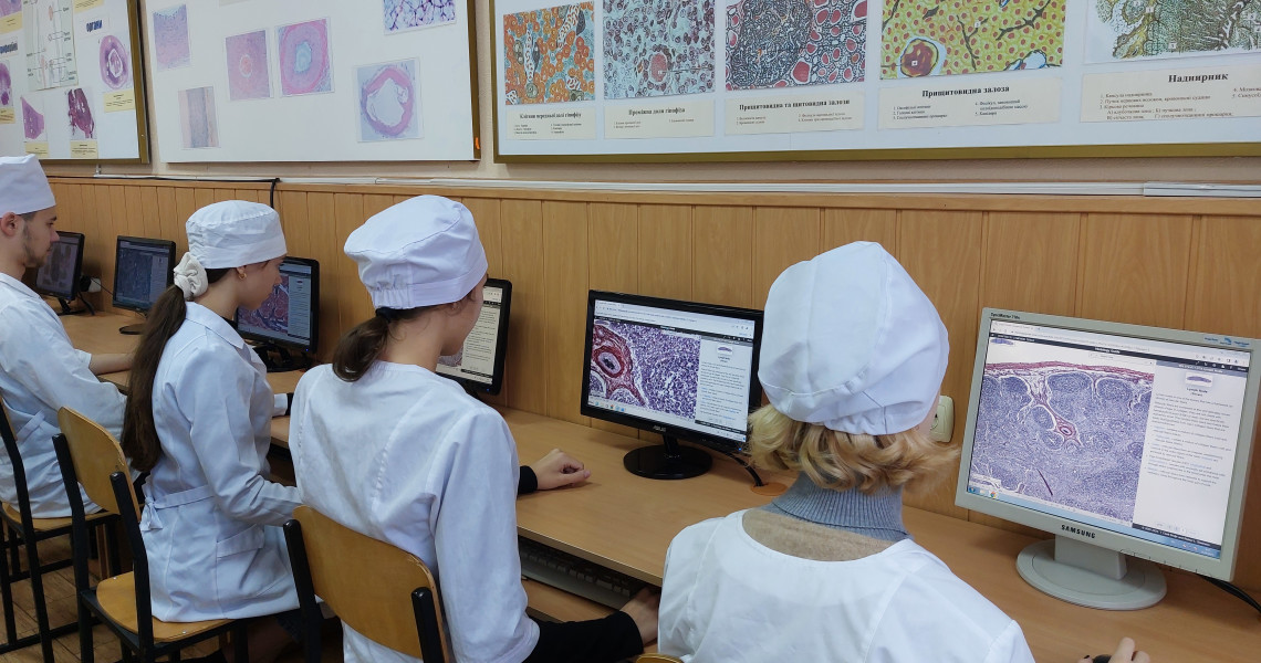
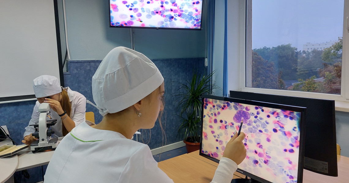
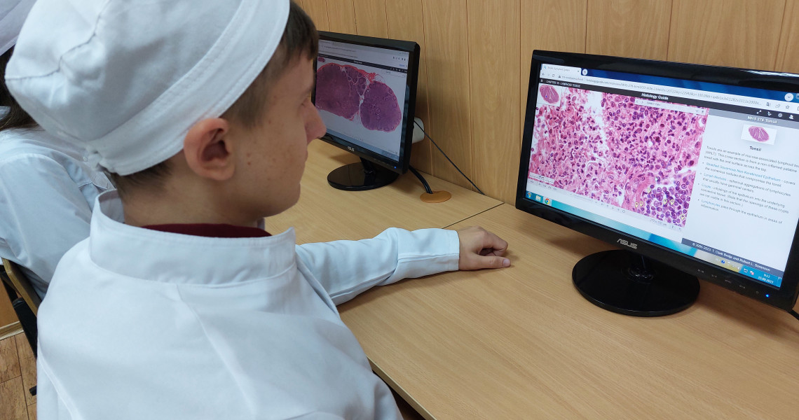
Другий етап полягає у візуалізації гістологічних препаратів за допомогою віртуальних гістологічних лабораторій “Histology Guide” та “Lumen Histology”. За допомогою даних програм гістологічні препарати вивчають, як на світлооптичному, так і електронномікроскопічному рівнях.
За допомогою новаторських технологій є можливість досліджувати тканини та органи при різних збільшеннях і методах забарвлення. При цьому здобувачі вищої освіти, перебувають у комфортних умовах кафедри та мають можливість засвоювати теоретичний матеріал порівнюючи схематичне зображення тканини чи органу з натуральним гістологічним препаратом.
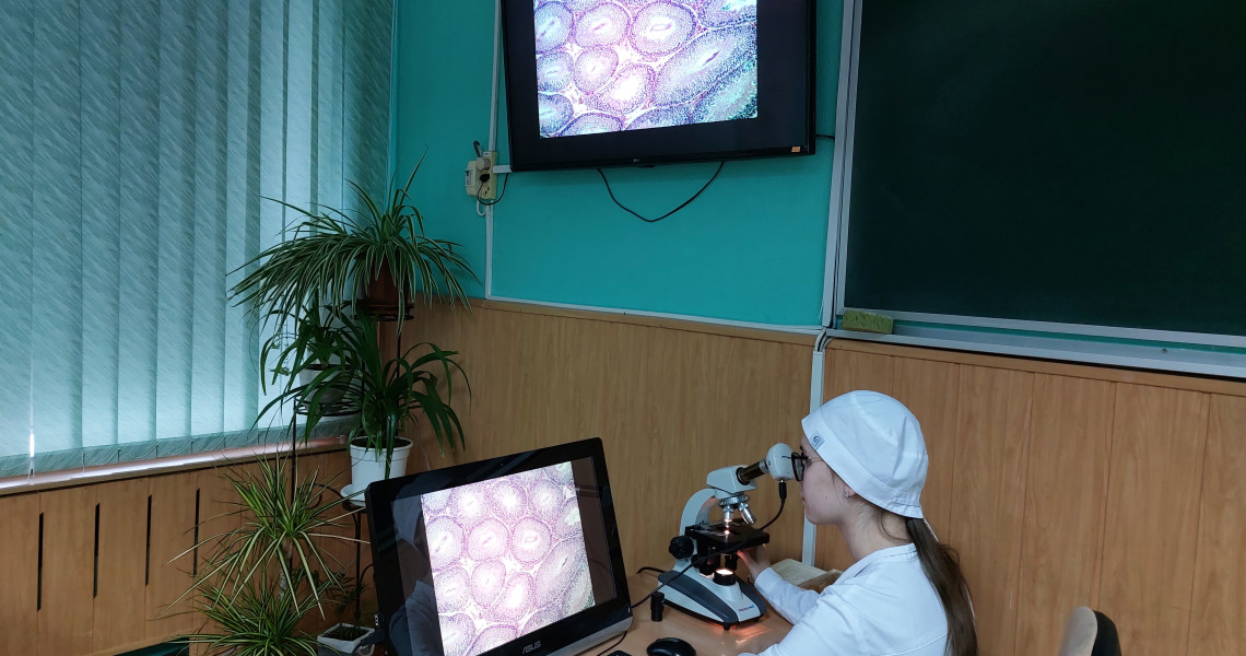
Під час третього етапу практичної частини заняття використовуються світлові мікроскопи. Здобувачі освіти самостійно, але під керівництвом викладача, вивчають мікропрепарати на предметному скельці.
Такий комплексний підхід до вивчення ОК «Гістологія, цитологія та ембріологія» оптимізує сприйняття мікроскопічної будови клітин, тканин і органів людини, яке вкрай необхідне для формування професійної компетентності наших здобувачів вищої освіти.
AN INNOVATIVE APPROACH TO THE ACQUISITION OF PRACTICAL SKILLS AT THE DEPARTMENT OF HISTOLOGY, CYTOLOGY, AND EMBRYOLOGY
The progress of medical science today is impressive, but without basic knowledge it is difficult to achieve success.
The obligatory component "Histology, Cytology and Embryology" is one of the basic morphological sciences. It is well known that the effectiveness of a future clinician depends on the diversity of his or her knowledge and skills. The practical classes therefore focus on the acquisition of practical skills, namely the study of the microscopic structure of cells and tissues through the diagnosis of histological specimens, which makes it possible to understand the rather complex spatial organisation of the human body.
In the practical lessons, the first step for the students is to work with images of the structural components of the organ at the light-optical level, followed by sketching them in compendiums and applying appropriate labels.
The second stage involves the visualisation of histological specimens using the Histology Guide and Lumen Histology virtual histology laboratories. With the help of these programmes, histological specimens are examined at both the light and electron microscopic levels.
Innovative technologies allow tissues and organs to be examined at various magnifications and staining methods. At the same time, students can stay in the comfortable conditions of the department and have the opportunity to learn theoretical material by comparing a schematic image of a tissue or organ with a natural histological specimen.
In the third stage of the practical part of the lesson, light microscopes are used. Students study microsections on a slide independently, but under the guidance of a teacher.
Such an integrated approach to the study of the EC 'Histology, Cytology and Embryology' optimises the perception of the microscopic structure of human cells, tissues and organs, which is essential for the development of our students' professional competence.


