


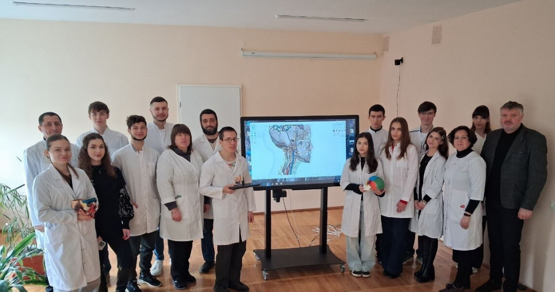
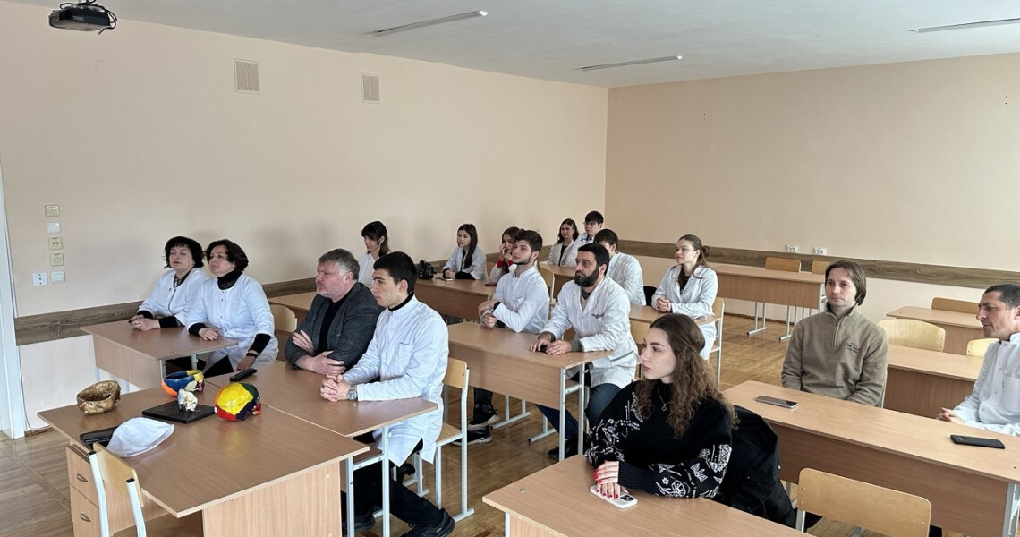
На кафедрі анатомії людини ПДМУ відбулося засідання студентської наукової групи, присвячене поглибленому вивченню топографічної анатомії черепа та інтеграції сучасних імерсивних технологій в освітній процес.
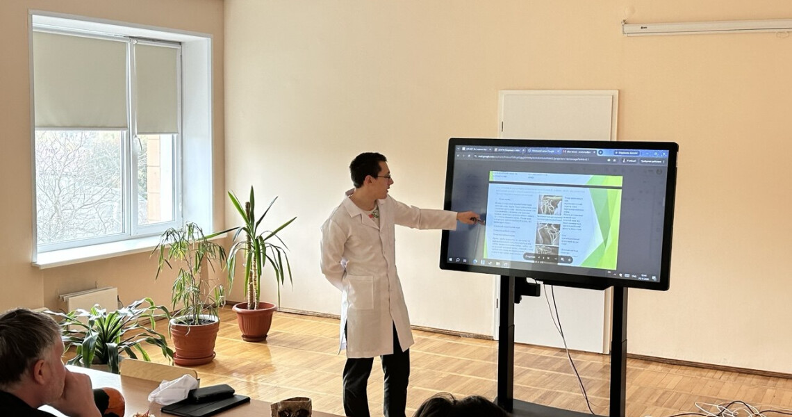
Під час засідання здобувач освіти ІІ курсу медичного факультету № 1 Сергій Тюрін презентував виготовлений ним анатомічний препарат «Крилопіднебінна ямка черепа». Під час демонстрації особливу увагу було приділено складній просторовій організації крилопіднебінної ямки, її анатомічним сполученням із порожниною черепа, очною ямкою, носовою та ротовою порожнинами, а також топографії крилопіднебінного вузла та його функціональному значенню. Учасники обговорили клінічну важливість знання анатомії цієї ділянки для стоматології, щелепно-лицевої хірургії та нейрохірургії.
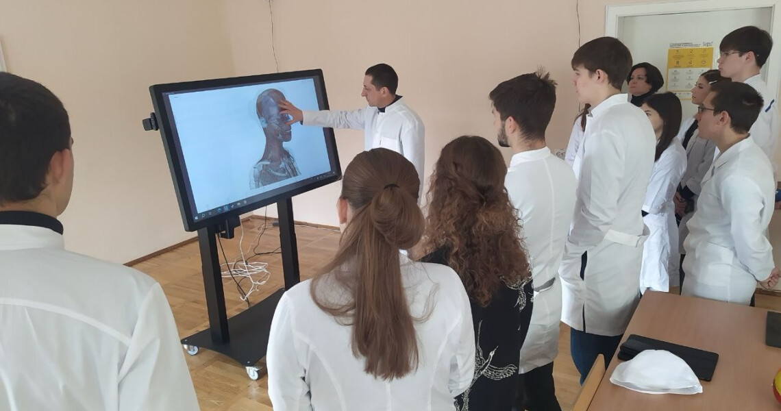
Практичну частину засідання провів доцент кафедри анатомії людини Андрій Пілюгін. У межах майстер-класу «Імерсивні технології в сучасній анатомічній освіті: від 3D-візуалізації до віртуальної реальності» він продемонстрував можливості інтерактивного анатомічного стола «Оніко».
Поєднання анатомічних препаратів із цифровими та імерсивними технологіями дає змогу краще вивчати просторове розташування анатомічних структур, наочно розглядати їхню будову та поглиблювати розуміння анатомії людини.
Пресслужба Полтавского державного медицинского университета
Anatomical Specimens and Immersive Technologies: Meeting of the Student Research Group at the PSMU Department of Human Anatomy
The Department of Human Anatomy of Poltava State Medical University (PSMU) hosted a meeting of its student research group dedicated to an in-depth study of the topographic anatomy of the skull and the integration of modern immersive technologies into the educational process.
During the meeting, Serhii Tiurin, a second-year student of Medical Faculty No. 1, presented an anatomical specimen he had prepared entitled “The Pterygopalatine Fossa of the Skull.” The demonstration focused particularly on the complex spatial arrangement of the pterygopalatine fossa, its anatomical communications with the cranial cavity, orbit, nasal and oral cavities, as well as the topography of the pterygopalatine ganglion and its functional significance. Participants discussed the clinical importance of understanding the anatomy of this region for dentistry, maxillofacial surgery and neurosurgery.
The practical part of the meeting was conducted by Andrii Piliuhin, Associate Professor of the Department of Human Anatomy. As part of the workshop entitled “Immersive Technologies in Modern Anatomical Education: From 3D Visualisation to Virtual Reality,” he demonstrated the capabilities of the ONIKO interactive anatomical table.
Combining anatomical specimens with digital and immersive technologies provides an opportunity to gain a better understanding of the spatial arrangement of anatomical structures, examine their morphology in detail and deepen knowledge of human anatomy.